Fertility NEWS LETTER
Ideal fertility : ICSI / IVF & Genetic Center India
Vol V Issue 6, June 2009
In this issue :
- IVF triplet pregnancy ends in HELLP syndrome with DIC
- A case of achondrogenesis.
- Ultrasound markers of successful pregnancy.
In previous issue :
- Chromosome abnormalities and Pregnancy loss
- Summer course on Biotechnology.
|
Dear Colleges
Hello
Hope this letter finds you in good health and good spirit.
In this issue I am discussing my two case I encountered in this month.
First was a nightmare for me . It was a case of HELLP syndrome with IVF triplet and with ? hepatic rupture. Patient collapsed on second post operative day but fortunately revived. It may be my mistake that I was not able to pre-empt the condition and be prepared for that. A preoperative platelet transfusion might have helped.( I need your comments on this case ,so that all our colleagues get benefited ).
But post operative HELLP syndrome is really very difficult case to manage, esp. in the days, when we doctors are frequently dragged in to the consumer redressal courts. Even a slight misdiagnosis(?) drags the attention of media and trial goes on in media.
One of my friend got in to this type of trouble and media exposed him like a culprit, without taking a plea that surgical complication is not always a professional negligence( until proved by the court).
This type of behaviour of media creates a big dent in the trust between the patients and the doctors.
Second case in an interesting rare case of ? achondrogenesis
An interesting finding in early pregnancy sonography marker for successful pregnancy is also presented.
Hope you will like these topics, Please feel free to give your comment on my choice of topics. Your suggestions will help to improve my News letter.
Bye
Sincerely Yours
Dr. D’Pankar Banerji
1. IVF triplet pregnancy ends in HELLP syndrome with DIC (disseminated intravascular coagulation)
Patient’s name is Usha (changed), aged 47 yrs.came to us for treatment from childless ness. She was perimenopausal with irregular periods. She has been advised for egg donation.
Two good quality blastocyst and one morula were transferred on 25th September 2008.
She conceived and three gestational sac with fetal cardiac activity were identified after one month. She refused embryo reduction.
The pregnancy was uneventful till April end. After that she started developing swelling in her feet. Her blood pressure and other parameters were within normal limits. Her blood pressure started rising from 3rd may 2009 to 150/98 mmHg. Tablet labetolol was started and she was monitored in hospital daily for BP and urine was examined for proteins, which was absent till end. It was treated as gestational hypertension and tab. Labetolol was continued. Steroid injections were given to mature the lungs of the babies. On 10th May she started swelling over face and upper limbs.
On 17th may she started leaking and an emergency Cesarean section was done undere GA. Three babies delivered.
Surgery was uneventful, but after one hour she started severe vaginal bleeding and her BP starts dropping. There was intra-abdominal blood (may be due to liver rupture). Her platelet went to 58,000/cc and SGPT was 55 IU Serum bilirubin 7.8 mg/dl. Her PT and APTT both were prolonged more than twice than control. FDPs were within limits. She was treated aggressively in ICU with multiple packed cell transfusions and platelet fractions and FFPs. A drain was put to remove the intra-abdominal blood ,2.5 liters of blood came out in first two days and then it continued for six days but reduced gradually.
Fortunately she survived the danger and doing well now.
Criteria for diagnosing HELLP (hemolysis, elevated liver enzyme and low platelets)
Hemolysis: Burr cells, schistocytes in blood smear, Bilirubin more than 1.2 mg/dl, absent plasma haptoglobin.
Elevated liver enzymes: AST > 72 IU/L, LDH >600
Low platelet count: Platelets < 100,000/cc
Approximately 70% of the cases oh HELLP occur in the ante-partum period with the rest occurring post- partum. The majority of cases (70%) occur between 28 and 36 weeks of gestation, 20 % occur after 37 weeks and 10 % before 26 weeks. The large majority (80%) of the postpartum cases occur within 48 hours of delivery.
HELLP is associated with 1% risk of maternal death. Most of these deaths are the consequence of abruptio placentae with disseminated intravascular coagulation, acute renal failure and pulmonary edema.
Hepatic rupture is a severe complication of women with HELLP syndrome. It may occur antepartum or postpartum, and in both cases the signs and symptoms are of profound circulatory collapse. The signs of peritoneal irritation and the progressive hypovolemia will point to intra-abdominal bleeding as the cause of the problem. The prognosis for preeclamptic patients with liver rupture is ominous.
Managing the HELLP syndrome requires aggressive approach. With packed cells, platelets and clotting factors (FFP) are very important.
Steroid may be an important to improve the clinical course; it may have a beneficial effect on recovering platelets.
Careful monitoring of their input/output, pulse oximetry and periodic auscultation of their lungs is all that is necessary to asses their pulmonary and renal situation.
2. Achondrogenesis:
This case was referred to us by one of our colleague. The couple has an earlier delivery of a male child with short limbs. The baby died after two days. The case was unreported and not diagnosed.
She again had a pregnancy and on USG scan at 18 weeks it was found that she is carrying a baby with very short limbs and there is no vertebral ossification centers.
As she had an earlier history of this type of child they were advised for termination of pregnancy and they agreed. This baby was with short limbs. A photograph was taken and a X-Ray was taken .
X-Ray revealed a typical features of short limbs. There is no ossification center of vertebrae and ribs are short .
A tentative diagnosis of achondrogenesis was done but yet to be confirmed by a geneticist.
Achondrognesis literally means ,there is lack of formation of the bone in all part of the body. It is also known as anosteogenesis, is a lethal chondrodystrophy characterized by extreme micromelia, a short trunk and macrocrania. The birth prevalence ranges from 0.09 to 0.23 in 10,000 births. It is of two types . More lethal form which is type I achondrogenesis and Type II ,the less severe form.
Type I is further subdivided into type IA and IB.
Prenatal diagnosis should be suspected on the basis of micromelia, lack of vertebral ossification and a large head with various degree of ossification of the calvarium. Polyhydramnios and hydrops have been associated with achondrogenesis.
Achondrogenesis types IA and IB are inherited in an autosomal-recessive pattern,whereas most cases of typeII and hypochondrogensis have been sporadic .
Primary defect in achondrogenesis type IA is unknown. The only gene known to be associated with achondrogenesis type IB is SLC26A2( DSDST) which encodes sulfate transporter protein. Because the basic defect in achondrogenesis type IB had been identified, diagnosis by molecular studey is possible. The distinction between achondrogenesis type IB( which has recurrence risk of 25% ) and the more frequent autosomal dominant achondrogenesis type II , which has a lower recurrence risk, is important for genetic counseling. A couple at risk of having a child with acondrogensis type IB may take advantage of the possiblilty of molecular prenatal diagnosis by CVS( chorionic villus sampling) or amniocentesis. Achondrogenesis type II/hypochondrognesis results from mutation in the COL2A1 gene. Prenatal diagnosis by molecular analysis of the COL2A1 gene is available.
COL2A1The official name of this gene is “collagen, type II, alpha 1.”
COL2A1 is the gene's official symbol. The COL2A1 gene provides instructions for making one component of type II collagen, called the pro-alpha1(II) chain. Type II collagen adds structure and strength to the connective tissues that support the body's muscles, joints, organs, and skin. Type II collagen is found primarily in cartilage, a tough but flexible tissue that makes up much of the skeleton during early development
SLC26A2The official name of this gene is “solute carrier family 26 (sulfate transporter), member 2.”
SLC26A2 is the gene's official symbol. The SLC26A2 gene provides instructions for making a protein that transports charged molecules (ions), particularly sulfate ions, across cell membranes. This protein appears to be active in many of the body's tissues, including developing cartilage. Cartilage is a tough, flexible tissue that makes up much of the skeleton during early development. Most cartilage is later converted to bone, except for the cartilage that continues to cover and protect the ends of bones and is present in the nose and external ears.
3. Ultrasound markers of successful pregnancy
Successful pregnancy can be predicted with up to 94% accuracy between days 33 and 36 using transvaginal ultrasound to detect 3 markers.
33-36 days post conception( approx.15-18 days of missing periods)
- Gestational sac diameter : 12mm and above
- Yolk sac diameter : 2-6 mm
- Presence of fetal cardiac activity
A study was conducted by lead author Soyoung Bae, MD, from the Department of Obstetrics/Gynecology, University of Toledo Medical Center in Ohio.( presented in 57th annual ACOG )
The study was designed as a case–control study of 1092 pregnancies, the majority of which were conceived with infertility treatments, so the precise date of conception could be known. The objective was to identify ultrasound markers to predict successful pregnancy in early pregnant patients.
For women older than 40 years, the rate of successful prediction was 82%. For women with a history of recurrent pregnancy loss, if all 3 markers were present, the rate of accurate prediction was 94%.
These markers are helpful to predict which pregnancies are going to end as miscarriage .
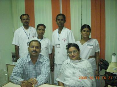
Our Staff :
| Archives |
- Vol VIII, Issue 11, Nov 2010
- Vol VIII, issue 6, June,2010
- Vol VIII,issue 5, May 2010
- Vol VIII,issue 4, April 2010
- Vol VIII Issue 3, March 2010
- Vol VIII, Issue 1,Jan 2010
- Vol VII, Issue 12,Dec.2009
- Vol VII, Issue 11,Nov.2009
- Vol VII, Issue 10,Oct.2009
- Vol VII, Issue 9, Sep.2009
- Vol VII, Issue 8, Aug 2009
- Vol VII, Issue 7,July 2009
- Vol VII, Issue 6,June 2009
- Vol VII Issue 4 april 2009
- Vol VI, Issue 9, Sep 2008
- Vol Vi Issue 8, aug 2008
- Vol Vi Issue 7, july 2008
- Vol VI, Issue 6, June 2008
- Vol V, Issue 17, may 2008
|
- Vol IV, Issue 16, April 2008
- Vol III, Issue 15, March 2008
- Vol I & II, Issue 13-14, Jan Feb 2008
- Vol IV, Issue 12, December 2007
- Vol IV, Issue 11, November 2007
- Vol IV, Issue 10, October 2007
- Vol IV, Issue 9, September 2007
- Vol IV, Issue 8, August 2007
- Vol IV, Issue 7, July 2007
- Vol IV, Issue 6, June 2007
- Vol IV, Issue 5, May 2007
- Vol IV, Issue 4, April 2007
- Vol IV, Issue 3, March 2007
- Vol IV, Issue 2, FEB_2007
- Vol IV, Issue1, Jan 2007
- Vol III, Issue 9, Nov Dec 2006
- Vol II, issue7, July 2005
- Vol II, Issue4 April 2005
- Vol II, Issue3, March 2005
|
|